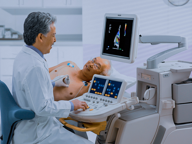
• 8:00am to 5:00pm Weekdays
  • (02) 9194 5000

Echocardiogram

An echocardiogram (or "echo") is a diagnostic test for the heart using ultrasound to produce live images of your heart. Echo uses standard two-dimensional, three-dimensional, and Doppler ultrasound technology to create images of the heart.

What should I expect?

An ultrasound is performed on the heart by applying an ultrasound wand (transducer) to the outside of the chest. Gel is applied to the chest to enable transmission of the ultrasonic waves. Detailed measurements of heart size and function and heart valve function are gathered. The test takes about 30 minutes and is performed whilst lying down. Three ECG electrodes are placed on your body whilst the images are being taken.

The test requires access to your chest area and you will be given a gown. It is recommended to wear two-piece attire.

A qualified cardiac sonographer will perform the examination. Images will be recorded and subsequently reviewed by the cardiologist. Your cardiologist will use this information to help with your overall cardiac treatment.

Results

The cardiologist and sonographer will create a report from your images, which will be sent electronically to your referring doctor. Please contact your referring doctor for your results and any necessary follow-up. If any significant abnormalities are detected, we will facilitate prompt treatment by contacting your referring doctor on the same day.
The information on this page is general in nature. All medical and surgical procedures have potential benefits and risks. Consult your medical practitioner for specific medical advice.
Experience excellence in cardiac care from leading cardiologists who place you at the centre of your care.
© 2025 Specialist Cardiology. All Rights Reserved.